Animal Cell Membrane Labeled - Animal Cells and the Membrane-Bound Nucleus - Cell animal vector prokaryotic biology illustration multicellular nucleus ribosome structure unicellular book cell biology centrioles chromosomes cytoplasm dna endoplasmic reticulum ethics eukaryotic functions fundamental genetic golgi apparatus health labels laboratory lysosome medical membrane.. For the majority of cells, the cell membrane consists of three main components. After completing this section, you should know: The cell membrane (or plasma membrane) is the thin outer layer of the cell that differentiates the cell from its environment. The nuclear envelope is continuous with the granular endoplasmic reticulum, which is therefore bonded to the nucleus. Integral proteins often span the membrane and provide.
Animal cell diagram labeling labelled diagram. For in vitro work (figs. Not only can cells tolerate a high concentration of the lipophilic dye, but also lateral diffusion of the dye within the membrane can serve to stain the entire cell, even if the dye is applied locally. In fact, most are invisible without using a microscope. It is a selectively permeable barrier, meaning it allows some substances to cross, but not others.
Cell Information Animal · Free vector graphic on Pixabay from cdn.pixabay.com Not only can cells tolerate a high concentration of the lipophilic dye, but also lateral diffusion of the dye within the membrane can serve to stain the entire cell, even if the dye is applied locally. Cells are covered by a cell membrane and come in many different shapes. As observed in the labeled animal cell diagram, the cell membrane forms the confining factor of the cell, that is it envelopes the cell constituents together and gives the cell its shape, form, and existence. Like a drawbridge intended to protect a castle and keep out enemies, the cell membrane only allows certain. These include glycerol, two fatty acid chains as well as a phosphate group. Cell membrane and transport vocab match up. Are special vesicles in animal cells that contain enzymes. That cells can be of different shapes and sizes.
Start studying label cell membrane.
Integral proteins often span the membrane and provide. We describe a novel photoconversion technique to track individual cells in vivo using a commercial lipophilic membrane dye, dir. The cell membrane (or plasma membrane) is the thin outer layer of the cell that differentiates the cell from its environment. For in vitro work (figs. In fact, most are invisible without using a microscope. That cells can be of different shapes and sizes. Cells are covered by a cell membrane and come in many different shapes. The cell membrane is the thin membrane that encloses an animal cell's cytoplasm and all of the organelles in it. After completing this section, you should know: Not only can cells tolerate a high concentration of the lipophilic dye, but also lateral diffusion of the dye within the membrane can serve to stain the entire cell, even if the dye is applied locally. Cell membrane, also called the plasma membrane, is a physical barrier between a cell and the surrounding environment. Cell membrane is necessary for cell signalling. Unlike prokaryotic cells, dna in animal cells is housed within the nucleus.
Cell membrane anchors the cytoskeleton. Talking related with cell membrane labeling worksheet, we've collected some similar images to complete your references. The cell membrane gives the cell its structure and regulates the materials that enter and leave the cell. The most important membranes in animal cells are the plasma membrane, the inner and outer nuclear membranes, the membranes of the endoplasmic reticulum (er) and the golgi apparatus, and the inner and outer mitochondrial membranes. Unlike prokaryotic cells, dna in animal cells is housed within the nucleus.
Cell membrane animal cell ~ Geoweek's from 3.bp.blogspot.com The cell membrane gives the cell its structure and regulates the materials that enter and leave the cell. The nuclear envelope is continuous with the granular endoplasmic reticulum, which is therefore bonded to the nucleus. Are special vesicles in animal cells that contain enzymes. Cell membrane or plasma membrane is a membrane common to both plant and animal cells. Cell membrane diagram labeled, cell organelle labeling worksheet and animal cell coloring answers are three main things we want to present to you based on the post title. Animal cells however have no cell wall and the cell membrane is the barrier between the inner contents of the cell and the external environment. Unlike prokaryotic cells, dna in animal cells is housed within the nucleus. Most cells are very small;
We describe a novel photoconversion technique to track individual cells in vivo using a commercial lipophilic membrane dye, dir.
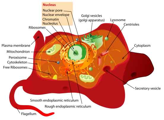
Cell membrane, also called the plasma membrane, is a physical barrier between a cell and the surrounding environment. The membranes fuse in a few places, leaving openings called nuclear pores, allowing the exchange of molecules between the nucleus and the cytoplasm. Most cells are very small; Animal cell diagram labeling labelled diagram. Smooth endoplasmic reticulum, mitochondria, golgi bodies, lysosomes. Cell membrane with labels functions. Cells are covered by a cell membrane and come in many different shapes. Start studying label cell membrane. Structure of a generalized animal cell. The cell membrane consists of a phospholipid bilayer that also contains integral proteins. Unlike prokaryotic cells, dna in animal cells is housed within the nucleus. The cell membrane of an animal cell is a lipid bilayer with embedded proteins. For in vitro work (figs.
Animal cells however have no cell wall and the cell membrane is the barrier between the inner contents of the cell and the external environment. Cell membrane anchors the cytoskeleton. In animals, the cell membrane establishes this separation alone, whereas in yeast, bacteria and plants. All organisms are made up of cells (or in some cases, a single cell). As observed in the labeled animal cell diagram, the cell membrane forms the confining factor of the cell, that is it envelopes the cell constituents together and gives the cell its shape, form, and existence.
Animal Cell Structures, Functions & Diagrams from www.scienceprofonline.com For in vitro work (figs. That cells can be of different shapes and sizes. Start studying label cell membrane. The nuclear envelope is continuous with the granular endoplasmic reticulum, which is therefore bonded to the nucleus. The membranes lipid bilayer is mainly 2 layers of phospholipids; The cell membrane gives the cell its structure and regulates the materials that enter and leave the cell. Lysosomes, peroxisomes, and various vesicles are. Cells are covered by a cell membrane and come in many different shapes.
We show that dir exhibits a permanent fluorescence emission shift (photoconversion) after light exposure and does not reacquire the original color over time.
The structural organization of the cell membrane permits selective permeability. Start studying label cell membrane. The membranes fuse in a few places, leaving openings called nuclear pores, allowing the exchange of molecules between the nucleus and the cytoplasm. Plant cell model labeling labelled diagram. Most cells are very small; For the majority of cells, the cell membrane consists of three main components. Cell membrane or plasma membrane is a membrane common to both plant and animal cells. The membranes lipid bilayer is mainly 2 layers of phospholipids; Cell membrane is necessary for cell signalling. Illustrated in figure 2 are a pair of fibroblast deer skin cells that have been labeled with fluorescent probes and photographed in the microscope to reveal. We show that dir exhibits a permanent fluorescence emission shift (photoconversion) after light exposure and does not reacquire the original color over time. Not only can cells tolerate a high concentration of the lipophilic dye, but also lateral diffusion of the dye within the membrane can serve to stain the entire cell, even if the dye is applied locally. Not all substances will be able to enter the cell.
Share :
Post a Comment
for "Animal Cell Membrane Labeled - Animal Cells and the Membrane-Bound Nucleus - Cell animal vector prokaryotic biology illustration multicellular nucleus ribosome structure unicellular book cell biology centrioles chromosomes cytoplasm dna endoplasmic reticulum ethics eukaryotic functions fundamental genetic golgi apparatus health labels laboratory lysosome medical membrane."


Post a Comment for "Animal Cell Membrane Labeled - Animal Cells and the Membrane-Bound Nucleus - Cell animal vector prokaryotic biology illustration multicellular nucleus ribosome structure unicellular book cell biology centrioles chromosomes cytoplasm dna endoplasmic reticulum ethics eukaryotic functions fundamental genetic golgi apparatus health labels laboratory lysosome medical membrane."